Practice with Real Ultrasound Data Training Simulator for Echocardiography and Point of Care Ultrasound (POCUS) in Neonates

Philosophy and Product Concept Concept
Echocardiography of the Newborn and Simulation
Echocardiography is the most important imaging technology to examine the neonatal heart. It is central in diagnosing congenital heart disease, one of the leading causes of neonatal mortality and morbidity in the developed world. Beyond that, examining the neonatal heart by echocardiography has emerged as essential in the management of sick neonates and preterm babies. One of the drawbacks of echocardiography is its user-dependency and its necessary high level of expertise. This expertise can only be achieved with considerable hands-on training. The delicate nature of neonates prohibits the acquisition of the necessary skills on sick patients. Simulation technology has increasingly become a standard in medical training. EchoCom|Neo now offers the opportunity to train echocardiography in neonates without putting patients at risk.
Our Philosophy
Echocom|Neo is the first ultrasound simulator worldwide dedicated to train echocardiography in infants and newborns. It is the result of 20 years of experience in simulation in echocardiography. The EchoCom simulator is based on real 3D ultrasound data. We believe that realistic data provide better acquisition of skills and knowledge than virtual models.
However, virtual models can help understand the 3D anatomy of the heart and its spatial relationship to the scan plane. We have therefore applied the concept of augmented reality that couples real and virtual data. A virtual probe and scan plane demonstrates this relationship through side-by-side presentation of the virtual and echocardiographic image. Echocom|Neo is designed to impart the two core skills needed by beginners: spatial sense and hand-eye-coordination.
Targeted Users
For the beginner in echocardiography data sets of normal hearts allow users to understand the complex anatomy of the heart, its visual presentation by echocardiography and to practice the movements required to obtain standard planes. The more advanced trainee can scan the database for typical neonatal cardiac problems and congenital heart diseases.
Point of Care Ultrasound Simulator
We have expanded EchoCom|Neo to include more organs. The echocardiography simulator thus matures to a full body ultrasound simulator.
Cerebral Ultrasound Simulation
The first organ that we implemented was the neonatal brain. As in the echocardiography module there are cases of normal cerebral ultrasound at different gestational ages as well as pathologies. And as in the cardiac module, real 3D ultrasound scans are used. It therefor allows you to explore the complex anatomical structure of the human brain.
Abdominal Ultrasound Simulation
Like the cardiac and cerebral modules real 3D ultrasound data is used. The size of the abdomen renders it impossible to have the abdomen in one 3D data set. The abdomen is therefore divided into different regions of interest. The presentation of umbilical vein catheters (with correct and incorrect position) should be of particular interest to neonatologists. A major problem poses the air filled bowel. The abdominal module is the least mature module, but as with the other organs, we work hard to overcome these obstacles.
Lung Ultrasound Simulation
The air filled lung poses special obstacles to a 3D simulation. Due to the air, lung ultrasound was considered useless or even impossible in the past. The ‚normal‘ lung itself cannot be represented abutting Thus, lung ultrasound is predominantly artifact-based, typically visualizing only abnormalities close to the pleural line. Generating a 3D ultrasound data set therefore is impossible. On the other hand the macroscopic appearance of the lung is quite simple. To generate a complex 3D model like in cardiac anatomy therefore is not necessary. Also probe handling is much more standardized and straightforward. Hand-eye-coordination is not as challenging as in echocardiography. The principle learning point in lung ultrasound simulation therefore is pattern recognition. Our approach to simulate lung ultrasound therefore is different form the previous modules. We use 2d video clips in longitudinal and transverse orientation from various positions to map the whole lung. A virtual baby demonstrates the location and orientation of each video clip.
Vascular Ultrasound Simulation
As in lung ultrasound hand-eye-coordination and mental 3D modelling of the organ are minor problems in vascular ultrasound. Demonstrating normal anatomy, technique of vascular access and possible complications are the major learning goals of this module.
We believe that this is a fantastic way to increase the exposure of neonatal and cardiology trainees to the whole range of structural abnormalities that newborn babies can have. For novice scanners in the NICU we think the simple act of getting your hands on the echo probe, getting used to seeing what a heart should look like, how the images change as you tilt and rotate the probe, will be a key foundation step in learning to perform echocardiography"
Dr. Alan Groves, Associate Professor, Department of Pediatrics, University of Texas at Austin, Texas, USA
Customers and Partners
Echocom|Neo is used by leading neonatal and cardiac medical centers, among others SickKids in Toronto, (the Canadian TnEcho National Collaborative), University of Iowa, USA (Prof. McNamara), Mount Sinai Hospital, New York, USA (Dr. Groves), Lucile Packard Children's Hospital Stanford, USA (Prof. Lopez), Children's Hospital of Philadelphia, USA (Dr. Manrique), the Rotunda Hospital, Dublin, Ireland (Dr. Al-Khuffash), University of Cambridge, UK (Dr. Singh), Radboud University Medical Center, Nijmegen, The Netherlands (Prof. de Boode) and University Hospital of North Tees, Durham University Stockton, UK (Dr. Gupta).
Echocom|Neo Product
Echocom|Neo consists of a dummy manikin and a dummy probe, a 3D tracking device and a computer with pre-installed software. The simulator fits in a custom made case to allow easy transport. On a split screen a virtual scene is presented side-by-side with a 2D echocardiograhic image derived from a 3D data set based on the tracked position of the dummy probe. Due to the use of real 3D data, the derived 2D image is highly realistic.
The images can be manipulated to adjust brightness, contrast, view standard planes or alter the heart rate. The virtual scene consists of a 3D heart that can be rendered transparent to see the valves, the thorax and a virtual ultrasound probe.
The manikin is a custom made life-sized newborn. The abdomen and neck are soft to allow indentation, while the thorax is made of less compressible material. The transmitter that is used for the tracking is incorporated into the manikin. A special silicon composition provides a highly realistic skin feel.
Echocom|Neo Library Library
The use of real 3D data enables us to build up a data base with a large number of case studies with all types of cardiac diseases. Our library encompasses data sets with the most common congenital cardiac defects and acquired heart diseases. Therefore it is suitable for physicians and technicians working in the field of pediatric cardiology and neonatologists performing echocardiography, so called targeted or functional neonatel echocardiography.
Examples of congenital heart disease cases
- Atrial septal defect
- Ventricular septal defect
- Atrioventricular septal defect
- Coarctation
- Tetralogy of Fallot
- Hypoplastic left heart syndrome
- Pulmonary atresia
- Tricuspid atresia
- Total anomalous pulmonary venous return
- Transposition of the great arteries
- Congenital corrected transposition of the great arteries
- Double outlet right ventricle
- Double inlet left ventricle
- Common arterial trunk
- Pulmonary stenosis
- Aortic stenosis
- Ebstein's anomaly
Examples of functional cases/acquired heart diseases
- Patent ductus arteriosus
- Persistent pulmonary hypertension of the newborn
- Pericardial effusion
- Cardiac tumors
- Hypertrophic cardiomyopathy
- Dilated cardiomyopathy
- Intracardiac thrombus
Examples of cerebral pathologies
- Normal neonatal brain
- Intraventricular hemorrhage
- Subarachnoideal hemorrhage
- Brain infarction
- Corpus callosum agenesis
- Dandy Walker malformation
- Hydrocephalus
The Echocom simulator is a state of the art interface which enhances the learning experience for trainees in the early part of their learning. In Canada we are now integrating the Echocom simulator into the curriculum of neonatal trainees who are learning Targeted Neonatal Echocardiography. It provides an accessible and feasible way to increase the trainees knowledge of congenital heart disease. We are most appreciative of the opportunity to use the simulator and thank Michael and Stefan for developing this for us. It is an incredible asset to neonatologists interested in learning echocardiography"
Prof. Patrick McNamara, Director, Division of Neonataology, University of Iowa, USA
Selected publications Publications
- Simulation-Based Echocardiography Training Enhanced Competence and Self-Confidence in Neonatal Residents. Blanchetière A, Guillouët E, Favrais G, Lardennois C, Bellot A, Savey B. Acta Paediatr. 2025 Jan
- Looking through the crystal ball. Feasibility of tele-echocardiography using smart glasses in neonates. Michaelis A, Weidenbach M, Altmann I, Markel F, Löffelbein F, Dähnert I, Gebauer RA, Paech C. Cardiol Young. 2025 Jan;35(1):87-92.
- Simulation in Neonatal Echocardiography. Weidenbach M, Paech C. Clin Perinatol. 2020 Sep;47(3):487-49
- Effectiveness of echocardiography simulation training for paediatric cardiology fellows in CHD. Dayton JD, Groves AM, Glickstein JS, Flynn PA. Cardiol Young. 2018 Jan 8:1-5
- Effectiveness of simulator based echocardiography training of non-cardiologists in congenital heart diseases. Wagner R, Razek V, Gräfe F, Berlage T, Janoušek J, Daehnert I, Weidenbach M. Echocardiography. 2013 Jul;30(6):693-8.
- Simulation of Congenital Heart Defects - A Novel Way of Training in Echocardiography. Weidenbach M, Razek V, Wild F, Khambadkone S, Berlage T, Janousek J, Marek J. Heart. 2009 Apr;95(8):636-41
- Computer Based Training in Two-dimensional Echocardiography Using an Echocardiography Simulator. Weidenbach M, Wild F, Scheer K, Muth G, Kreutter S, Grunst G, Berlage T, Schneider P. J Am Soc Echocardiogr. 2005; 18(4): 362-366
- Augmented Reality Simulator for Training in Two-Dimensional Echocardiography. Weidenbach M, Wick C, Pieper S, Quast KJ, Fox T, Grunst G, Redel DA. Comp Biomed Res. 2000 Feb; 33(1): 11-22
Training could also be augmented by use of simulators in neonatal echocardiography that are increasingly used for teaching on echocardiography courses. This could help in developing skills in acquiring standard echocardiographic views and recognizing common congenital heart conditions.”
Expert consensus statement ‘Neonatologist-performed Echocardiography (NoPE)’—training and accreditation in UK. Singh-Y et al. EurJPediatr(2016)175:281–287
News News
The use of simulators for training in neonatologist performed echocardiography is now endorsed by the European Society for Neonatology.
Read more…
Newsletter
Contact the Company Contact
For more specifics please contact us
EchoCom|NEO Support Support
The current software version of EchoCom|NEO is 1.3.1
For existing customers, please contact us at support@echocom.de or use the contact form.
We will respond as soon as possible!
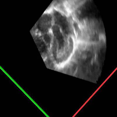 Apical four to five chamber sweep, normal heart
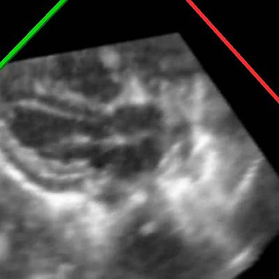 Parasternal long axis view, normal heart
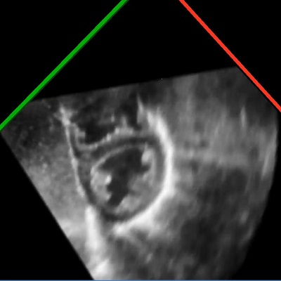 parasternal short axis, sweep from apex to the base, normal heart
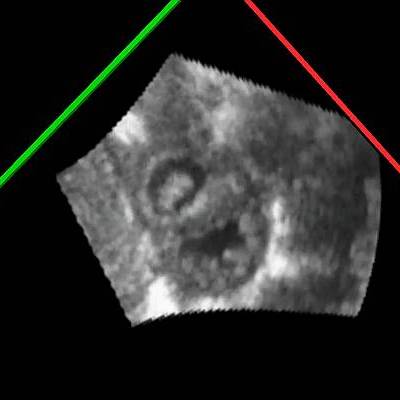 Persistent Pulmonary Hypertension of the Newborn (PPHN), parasternal short axis view
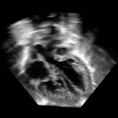 Transposition of the great arteries, subcostal view
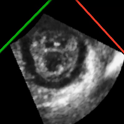 Pericardial effusion
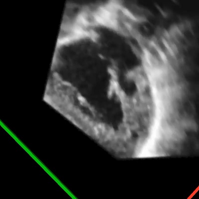 Hypoplastic left heart syndrome, apical view
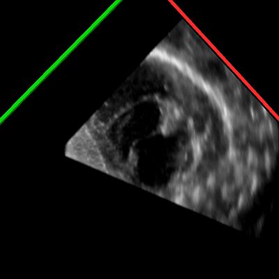 PDA, ductal view
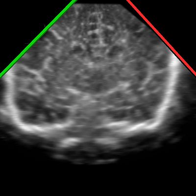 Normal cranial ultrasound
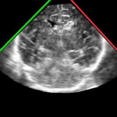 Intraventricular Hemorrhage Grade-2 left side, Grade-1 right side
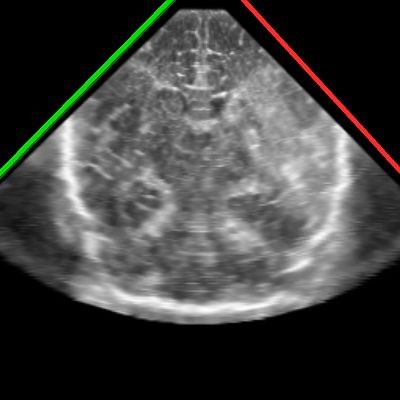 Middle Cerebral Artery Infarct, left side
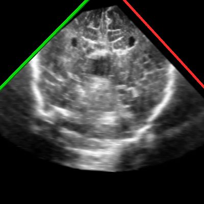 Corpus Callosum Agenesis